Cell and Molecular

Mitochondria: Introduction to Cell Communication

Learning Objectives

  • Understand the Structure and Function of Mitochondria
  • Mitochondria and Cellular Signaling
  • Mitochondrial Dysfunctions and Disease
  • Cell Communication

Mitochondria

Small organelles in eukaryotic cells often referred to as the “powerhouse of the cell.” They generate ATP through oxidative phosphorylation, contain their own DNA, and are believed to have originated from endosymbiotic bacteria.

Structure

  • Outer Membrane: A simple phospholipid bilayer containing large integral proteins called porins. It allows the passage of ions, nutrient molecules, ATP, ADP, etc., with ease.
  • Inner Membrane: Freely permeable only to oxygen, CO2, and H2O. It contains proteins for oxidative phosphorylation, ATP synthase, transport proteins, and machinery for mitochondrial fusion and fission.
  • Intermembrane Space: Known as the perimitochondrial space, it has a high proton concentration.
  • Matrix: Enclosed by the inner membrane, this space contains enzymes, mitochondrial DNA, ribosomes, tRNA, and other components.
  • Cristae: Folds of the inner membrane that increase surface area, enhancing ATP production.

Functions of Mitochondria

Primary Role: Production of ATP through cellular respiration.

Other Functions:

  • Regulation of metabolic pathways
  • Calcium signaling
  • Apoptosis (programmed cell death)
  • Heat production (thermogenesis)

Introduction to Cell Communication

Cell communication is the process by which cells detect and respond to signals in their environment. It involves chemical signals like hormones, neurotransmitters, and other molecules.

This process is crucial for maintaining homeostasis, coordinating cell activities, and responding to external stimuli.
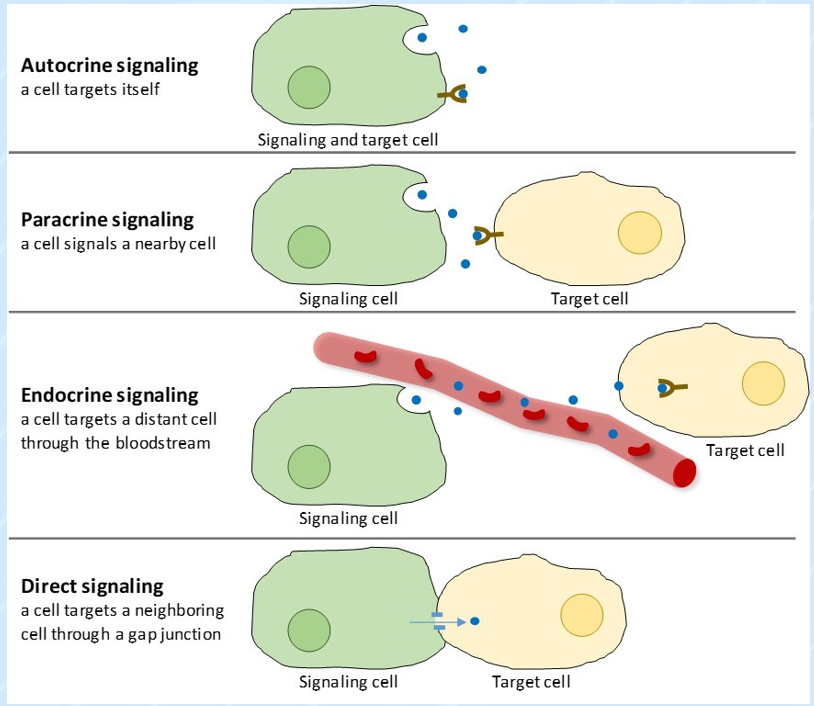
Forms of Signaling

  • Autocrine signaling
  • Paracrine signaling
  • Endocrine signaling
  • Direct contact

Mitochondria and Cell Signaling

Mitochondria play a key role in regulating important cell signaling pathways, including:

  • Calcium Signaling: Mitochondria act as calcium buffers, regulating intracellular calcium levels and preventing calcium overload.
  • Reactive Oxygen Species (ROS) Signaling: ROS are byproducts of mitochondrial activity. Low levels act as signaling molecules, but excessive ROS cause oxidative stress, leading to cellular damage.
  • Apoptosis: Mitochondria are critical in initiating programmed cell death, which removes damaged or unwanted cells.

Mitochondrial Dysfunction and Cell Communication

Mitochondrial dysfunction impairs cellular signaling, leading to issues like cell death, uncontrolled growth, or failure to respond to environmental signals. Conditions linked to dysfunction include:

  • Neurodegenerative diseases (e.g., Alzheimer’s, Parkinson’s)
  • Oxidative stress and inflammation
  • Calcium overload

Mitochondrial Diseases

  • Mitochondrial encephalopathy, lactic acidosis, and stroke-like episodes (MELAS)
  • Leigh syndrome
  • Leber hereditary optic neuropathy (LHON)
  • Kearns-Sayre syndrome (KSS)
  • Myoclonic epilepsy and ragged-red fiber disease (MERRF)

Neurodegenerative Diseases

  • In these diseases, abnormal mitochondrial function leads to excessive ROS production and defective ATP generation, contributing to neuron death.
  • Impaired mitochondrial dynamics, including fusion and fission processes, exacerbate the disease progression.

Mitochondria in Cancer

  • Cancer cells often exhibit altered mitochondrial function, promoting aerobic glycolysis (Warburg effect) even in the presence of oxygen.
  • Mitochondria support cancer cell survival and proliferation by controlling ROS levels, promoting resistance to apoptosis, and contributing to tumor metastasis.
  • Targeting mitochondrial function is a promising strategy in cancer therapy.

Mitochondrial dysfunction contributes to neurodegenerative diseases, cancer, and other conditions by promoting oxidative damage and affecting ATP production.

Cellular Tensegrity and Molecular Motors

Introduction

Understanding Structural Integrity and Force Generation in Cells

  • Structural integrity: Refers to cells' ability to maintain shape and resist deformation, ensuring they don’t collapse under stress.
  • Microfilaments (actin filaments): Provide tension and resistance to pulling forces.
  • Microtubules: Act like compression rods, resisting compression and aiding in cell transport and division.
  • Intermediate filaments: Provide structural support and durability, especially in cells under constant stress, like skin cells.
  • Force generation: Necessary for cell movement, division, and interactions. Examples include muscle contractions, cell migration, and tissue development.
  • Actin-myosin interactions: Actin filaments and myosin motor proteins create tension and generate force.
  • Cell migration: Actin polymerization allows cells to extend protrusions (e.g., lamellipodia and filopodia) to push the cell membrane forward.

Introduction to Cellular Mechanics

Cellular mechanics are essential for biological systems' functionality. The structure of a cell helps maintain its shape and enables specific functions. External forces (pressure, tension, or compression) significantly impact physiology and behavior as cells adjust their internal structure and activity in response.

What is Tensegrity?

Tensegrity: A design principle where a structure maintains its shape through a balance of tensile (pulling) and compressive (pushing) forces.

Importance in biology: Tensegrity explains how cells and tissues maintain shape and handle forces. The cytoskeleton acts like a tensegrity structure, where interconnected filaments manage tension and compression for stability and flexibility.

In signal transduction, tensegrity enables cells to sense and respond to mechanical cues. External forces alter cytoskeletal tension, triggering cellular signals.

Introduction to Molecular Motors

Molecular motors: Proteins functioning as tiny machines within cells. They convert chemical energy (from ATP) into mechanical work to drive cellular processes.

  • Myosin: Involved in muscle contraction and cell movement.
  • Kinesin: Transports cargo (e.g., proteins or organelles) along microtubules outward from the cell center.
  • Dynein: Moves cargo toward the cell center.

Mechanisms of Molecular Motors

  1. ATP Hydrolysis: Motor proteins bind ATP, breaking it down into ADP and inorganic phosphate, releasing energy.
  2. Conformational Changes: Energy from ATP hydrolysis alters motor protein shape.
  3. Mechanical Work: Proteins move along filaments, transporting cargo.

Molecular motors and tensegrity collaborate to maintain structural stability, generate force, and support dynamic cellular functions.

Tensegrity and Molecular Motors Interactions

  • Force generation and distribution: Molecular motors act on the cytoskeleton, reinforcing tensegrity.
  • Cell shape changes: Motors pull on cytoskeletal filaments, enabling cell contraction, spreading, or movement.
  • Mechanotransduction: Tensegrity and motor-generated forces help cells sense and respond to mechanical signals.
  • Intracellular transport: Motors transport organelles and molecules along cytoskeletal tracks, ensuring efficient material delivery.

Cellular Tensegrity and Disease

  • Cancer metastasis: Disruptions in tension and cytoskeletal structure aid tumor cell shape changes and migration.
  • Cardiovascular diseases: Altered tension impacts contractility and elasticity, affecting vessel and heart muscle function.
  • Muscular dystrophies: Defective tension regulation, often due to cytoskeletal protein mutations.

Therapeutic implications: Targeting cellular mechanics offers treatment potential. Drugs modulating tension or motor protein function could limit cancer spread, improve heart function, or stabilize muscle cells in muscular dystrophy.

Watch the presentation video: Click here

Cell Adhesion and Cell Junction

What is Cell Adhesion?

The process by which cells attach to each other or to the extracellular matrix.

What is Extracellular Matrix?

A network of proteins and carbohydrates that surrounds cells, providing structural support and influencing cell behavior.

What is a Cell Junction?

Specialized structures that connect cells to each other or to the extracellular matrix (ECM).

Composition of Cell Junctions

  • Proteins:
    • Transmembrane proteins: Cadherins, integrins, claudins, occludins
    • Cytoplasmic adaptor proteins: Catenins, plakoglobin, vinculin
  • Cytoskeleton components: Actin filaments or intermediate filaments

Functions of Cell Junctions

  • Adhesion
  • Communication
  • Barrier Formation
  • Signal Transduction
  • Cell Polarity

Types of Cell Junctions (Animal Cells)

1. Tight Junctions (Occluding Junctions)

Composition:

  • Transmembrane proteins
  • Cytoplasmic adaptor proteins
  • Linked to actin cytoskeleton

Functions:

  • Prevent leakage of molecules between cells
  • Maintain cell polarity by separating apical and basolateral surfaces
  • Act as a barrier to pathogens and toxins

Example: Small intestine epithelium prevents leakage of digestive enzymes and pathogens into the bloodstream.

2. Adherens Junctions

Composition:

  • Transmembrane proteins
  • Cytoplasmic adaptor proteins
  • Linked to actin filaments

Functions:

  • Provide mechanical adhesion between cells
  • Facilitate coordinated cell movement during tissue remodeling

Example: Skin epidermis provides strength to resist mechanical stress from external forces.

3. Desmosomes (Anchoring Junctions)

Composition:

  • Transmembrane proteins
  • Cytoplasmic adaptor proteins
  • Linked to intermediate filaments (keratin)

Functions:

  • Strengthen intercellular connections
  • Resist mechanical stress by distributing tension

Example: Heart muscle cells maintain structural integrity during constant contraction and relaxation.

4. Gap Junctions (Communicating Junctions)

Composition:

  • Transmembrane proteins

Functions:

  • Allow direct transfer of ions, nutrients, and signaling molecules between cells
  • Enable electrical and chemical communication

Example: Cardiac muscle cells facilitate synchronized contraction by allowing ion flow between cells.

5. Hemidesmosomes

Composition:

  • Transmembrane proteins
  • Linked to intermediate filaments (keratin)

Functions:

  • Anchor cells to the extracellular matrix (e.g., basement membrane)
  • Provide resistance to shear forces

Example: The basal layer of skin epidermis keeps skin attached to the underlying dermis, ensuring durability under friction.

Type of Junction Composition Functions Example
Tight Junctions (Occluding Junctions) - Transmembrane proteins
- Cytoplasmic adaptor proteins
- Linked to actin cytoskeleton
- Prevent leakage of molecules between cells
- Maintain cell polarity by separating apical and basolateral surfaces
- Act as a barrier to pathogens and toxins
Small intestine epithelium prevents leakage of digestive enzymes and pathogens into the bloodstream.
Adherens Junctions - Transmembrane proteins
- Cytoplasmic adaptor proteins
- Linked to actin filaments
- Provide mechanical adhesion between cells
- Facilitate coordinated cell movement during tissue remodeling
Skin epidermis provides strength to resist mechanical stress from external forces.
Desmosomes (Anchoring Junctions) - Transmembrane proteins
- Cytoplasmic adaptor proteins
- Linked to intermediate filaments (keratin)
- Strengthen intercellular connections
- Resist mechanical stress by distributing tension
Heart muscle cells maintain structural integrity during constant contraction and relaxation.
Gap Junctions (Communicating Junctions) - Transmembrane proteins - Allow direct transfer of ions, nutrients, and signaling molecules between cells
- Enable electrical and chemical communication
Cardiac muscle cells facilitate synchronized contraction by allowing ion flow between cells.
Hemidesmosomes - Transmembrane proteins
- Linked to intermediate filaments (keratin)
- Anchor cells to the extracellular matrix (e.g., basement membrane)
- Provide resistance to shear forces
The basal layer of skin epidermis keeps skin attached to the underlying dermis, ensuring durability under friction.

Types of Cell Junctions (Plant Cells)

Plasmodesmata

Composition:

  • Plasma membrane
  • Cytoplasmic sleeve
  • Desmotubules
  • Proteins and molecules

Functions:

  • Transport: Move small molecules like water, sugars, amino acids, and ions between cells
  • Signal Transduction: Allow movement of signaling molecules and hormones to coordinate cellular activities
  • Integration: Enable plant cells to work together as a unified tissue for growth, repair, and defense
  • Developmental Control: Regulate the movement of transcription factors and RNA during growth and differentiation

Example: Found in leaf mesophyll cells. They distribute sugars made during photosynthesis to nearby cells for storage or transport and facilitate efficient communication to regulate stomatal opening and closing in response to environmental changes.

Connective Tissue

Stem Cells and Engineering

Learning Objectives

  • Identify the three types of connective tissue
  • Compare and contrast each type and their variations
  • Demonstrate how connective tissue works together

Connective Tissue

These tissues connect and support other tissues of the body.

Functions:

  • Mechanical support
  • Medium for exchange of nutrients & waste products
  • Energy store and thermal insulation
  • Defensive functions:
    • Barrier
    • Engulf bacteria
    • Antibodies

Composition of Connective Tissue

  • Fibroblasts
  • Plasma Cells
  • Adipose Cells
  • Large Lymphocytes
  • Macrophages
  • Fibrocytes
  • Eosinophils
  • Neutrophils
  • Cells with pigment granules
  • Small Lymphocytes
  • Mast Cells

Fibers:

  • Collagen Fibers: Large fibers made of collagen, promoting tissue flexibility.
  • Elastic Fibers: Intermediate fibers made of elastin, allowing for stretch and recoil.
  • Reticular Fibers: Small, delicate, branched fibers forming structural frameworks for organs like the spleen and lymph nodes.

Classification of Connective Tissue

Loose Connective Tissue:

  • Areolar Connective Tissue: Forms a loose network in intracellular material, providing strength, elasticity, and support. Found beneath the skin and around blood vessels and nerves.
  • Adipose Connective Tissue: Contains adipocytes that store fat, providing energy storage, heat insulation, and protection for organs. Found in the subcutaneous layer, around the heart, and kidneys.
  • Reticular Connective Tissue: Contains reticular fibers and cells that form a supportive framework for organs like the liver, spleen, and lymph nodes. Also involved in filtering microbes in lymph nodes.

Dense Connective Tissue:

  • Dense Regular Connective Tissue: Collagen fibers are arranged in parallel patterns, providing strong attachments (e.g., tendons and ligaments).
  • Dense Irregular Connective Tissue: Irregularly arranged collagen fibers provide strength to the dermis and organ capsules (e.g., around kidneys and liver).
  • Elastic Connective Tissue: Contains freely branching elastic fibers, allowing stretch and recoil (e.g., in arteries and vocal cords).

Cartilage:

  • Hyaline Cartilage: Bluish-white cartilage providing smooth surfaces for joint movements. Found in ribs, nose, and respiratory passages.
  • Fibrocartilage: The strongest cartilage, containing dense bundles of collagen. Found in intervertebral discs, providing support and shock absorption.
  • Elastic Cartilage: Contains elastic fibers for flexibility, maintaining the shape of organs like the ear and epiglottis.

Other Types:

  • Bone Tissue: Provides structural support and protects organs.
  • Liquid Connective Tissue: Blood and lymph transport nutrients, gases, and waste products.

Stem Cells and Tissue Engineering

An interdisciplinary field combining biology, medicine, and engineering to design functional components for tissue maintenance, replacement, or regeneration.

Stem Cells:

Undifferentiated cells capable of self-renewal and differentiation into various cell types during development and adulthood.

  • Embryonic Stem Cells: Pluripotent with high self-renewal capacity but pose ethical challenges.
  • Adult Stem Cells: Found in most tissues; less controversial but limited in differentiation capacity.
  • Induced Pluripotent Stem Cells (iPSCs): Reprogrammed adult cells with capabilities similar to embryonic stem cells.

Regeneration and Tissue Engineering

Exploring regeneration possibilities in humans with examples from nature like planarians, crayfish, and embryos. The complexity of organisms inversely relates to regenerative ability.

Models for Tissue Engineering:

  • Injectable stem cells for minimally invasive procedures
  • Solid scaffold manufacturing for custom tissue constructs

Thank you for your attention! I hope you learned something new today!

1. Mitochondria: Introduction to Cell Communication

Mitochondria

Definition:
Small organelles in eukaryotic cells. Known as the “powerhouse of the cell” for generating ATP. Contains its own DNA; evolved from endosymbiotic bacteria.

Structure

  • Outer Membrane: Contains porins allowing the passage of ions, ATP, ADP, etc.
  • Inner Membrane: Permeable to Oxygen, CO2, H2O. Houses proteins for oxidative phosphorylation and ATP production.
  • Intermembrane Space: High proton concentration.
  • Matrix: Contains enzymes, DNA, ribosomes, and tubules.
  • Cristae: Folds in the inner membrane, increasing the surface area for ATP production.

Functions of Mitochondria

Primary Role:
ATP production through cellular respiration.

Other Functions:

  • Regulation of metabolic pathways
  • Calcium signaling
  • Apoptosis (programmed cell death)
  • Heat production (thermogenesis)

Cell Communication

Definition:
Process by which cells detect and respond to signals in their environment.

Forms of Signaling

  • Autocrine signaling: Cell signals itself.
  • Paracrine signaling: Signals nearby cells.
  • Endocrine signaling: Signals travel through the bloodstream.
  • Direct contact: Physical interaction between cells.

Mitochondria and Cell Signaling

  • Calcium Signaling:
    Regulates intracellular calcium levels, affecting muscle contraction, neurotransmitter release, and enzyme activity. Disruption leads to diseases like neurodegeneration.
  • Reactive Oxygen Species (ROS) Signaling:
    Low ROS: Acts as a signaling molecule for growth and immune responses.
    High ROS: Causes oxidative stress, damaging DNA, proteins, and lipids.
  • Apoptosis:
    Mitochondria release cytochrome c, activating enzymes for cell death. Essential for eliminating damaged cells.

Mitochondrial Dysfunction

Impact on Cell Communication:
Disrupts:

  • Calcium regulation
  • ROS signaling
  • Cell response to environmental signals

Linked Diseases:

  • Neurodegenerative diseases: Alzheimer’s, Parkinson’s
  • Cardiovascular diseases: From inflammation and oxidative stress

Mitochondrial Diseases

  • MELAS: Stroke-like episodes
  • Leigh syndrome: Neurological disorder
  • LHON: Vision loss
  • Kearns-Sayre syndrome (KSS): Affects muscle and eye movement
  • MERRF: Myoclonic epilepsy

Mitochondria in Cancer:
Cancer cells alter mitochondrial function, promoting the Warburg effect (glycolysis even with oxygen present). This supports tumor growth by regulating ROS and preventing apoptosis.

Key Takeaways

  • Mitochondria are essential for energy production, calcium signaling, and cell death regulation.
  • Disruptions in mitochondrial function can lead to serious diseases, including cancer and neurodegenerative conditions.
  • Understanding mitochondrial behavior provides insight into potential therapeutic approaches for many diseases.

Study Tip: Use this as a summary sheet and refer back to your materials for detailed explanations on each topic!

2. Study Material: Cell Adhesion and Junctions

Cell Adhesion

Definition: The process by which cells attach to each other or to the extracellular matrix (ECM).

Extracellular Matrix (ECM)

Definition: A network of proteins and carbohydrates that surrounds cells, providing structural support and influencing cell behavior.

Cell Junctions

Definition: Specialized structures that connect cells to each other or to the ECM.

Key Functions:

  • Adhesion: Ensures cells stay connected.
  • Communication: Allows exchange of signals and materials.
  • Barrier Formation: Prevents unwanted movement of substances.
  • Signal Transduction: Facilitates responses to environmental signals.
  • Cell Polarity: Maintains distinct cellular orientations.

Types of Cell Junctions in Animal Cells

Type Key Composition Key Functions Example in Function
Tight Junctions Transmembrane proteins, adaptor proteins, actin cytoskeleton
  • Prevent leakage of molecules between cells.
  • Maintain cell polarity by separating surfaces.
Small intestine epithelium: Prevents digestive enzyme and pathogen leakage.
Adherens Junctions Transmembrane proteins, adaptor proteins, actin filaments
  • Provide mechanical adhesion.
  • Facilitate coordinated cell movement during tissue remodeling.
Skin epidermis: Resists mechanical stress.
Desmosomes Transmembrane proteins, adaptor proteins, intermediate filaments
  • Strengthen intercellular connections.
  • Distribute mechanical stress.
Heart muscle cells: Maintain structural integrity during contraction.
Gap Junctions Transmembrane proteins
  • Allow direct transfer of ions and molecules between cells.
  • Enable electrical and chemical communication.
Cardiac muscle cells: Facilitate synchronized contraction by allowing ion flow.
Hemidesmosomes Transmembrane proteins, intermediate filaments
  • Anchor cells to ECM (e.g., basement membrane).
  • Provide resistance to shear forces.
Basal layer of skin epidermis: Maintains attachment to underlying dermis.

Types of Cell Junctions in Plant Cells

Type Key Composition Key Functions Example in Function
Plasmodesmata Plasma membrane, cytoplasmic sleeve, desmotubules
  • Transport small molecules (water, sugars, amino acids, ions).
  • Coordinate cellular activities through signal transduction.
Leaf mesophyll cells: Distribute sugars made during photosynthesis.

Distinct Characteristics of Junctions

  • Tight Junctions: Create impermeable barriers; maintain polarity in epithelial cells.
  • Adherens Junctions: Connect cells mechanically via actin cytoskeleton; key during tissue remodeling.
  • Desmosomes: Provide strong adhesion through keratin filaments; critical in mechanically stressed tissues.
  • Gap Junctions: Directly link cytoplasms for communication; enable synchronization in muscles.
  • Hemidesmosomes: Anchor cells to ECM; essential for epithelial structural stability.
  • Plasmodesmata (Plants): Allow integrated function and signaling across plant tissues.

3. Study Material: Cellular Tensegrity and Molecular Motors

Introduction

Structural Integrity in Cells:

  • Maintains shape and resists deformation.
  • Key Components:
    • Microfilaments (Actin Filaments): Resist pulling forces.
    • Microtubules: Resist compression, assist in transport and division.
    • Intermediate Filaments: Provide durability in stress-prone cells (e.g., skin cells).

Force Generation in Cells:

  • Necessary for movement, division, and interaction.
  • Key Processes:
    • Actin-Myosin Interactions: Create tension and generate force.
    • Cell Migration: Actin polymerization extends protrusions (lamellipodia, filopodia) to push the membrane forward.

Cellular Mechanics

  • Essential for:
    • Shape Maintenance and Functionality.
    • Response to external forces (pressure, tension, compression).
  • Cells adjust their internal structure and activity in response to mechanical stimuli.

Tensegrity in Biology

Definition: A balance of tensile (pulling) and compressive (pushing) forces maintaining structural stability.

Role in Biology:

  • The cytoskeleton acts as a tensegrity structure, balancing tension and compression.
  • Signal Transduction: External forces alter cytoskeletal tension, triggering cellular signals.

Molecular Motors

Definition: Proteins that convert ATP (chemical energy) into mechanical work for cellular processes.

Main Types:

  • Myosin: Involved in muscle contraction and cell movement.
  • Kinesin: Transports cargo outward along microtubules.
  • Dynein: Transports cargo inward toward the cell center.

Mechanisms of Molecular Motors

  1. ATP Hydrolysis: ATP is broken into ADP and inorganic phosphate, releasing energy.
  2. Conformational Changes: Energy from ATP hydrolysis changes motor protein shape.
  3. Mechanical Work: Motor proteins move along filaments, transporting cellular cargo.

Molecular motors and tensegrity collaborate to maintain structural stability, generate force, and support dynamic cellular functions.

Interactions Between Tensegrity and Molecular Motors

  • Force Generation and Distribution: Molecular motors reinforce cytoskeleton tensegrity by generating forces.
  • Cell Shape Changes: Motors pull on cytoskeletal filaments, enabling contraction, spreading, and movement.
  • Mechanotransduction: Combined tensegrity and motor forces allow cells to sense and respond to mechanical signals.
  • Intracellular Transport: Efficient transport of organelles and molecules along cytoskeletal "tracks."

Cellular Tensegrity and Disease

Key Diseases Linked to Cellular Tensegrity:

  • Cancer Metastasis: Disruptions in tension and structure facilitate tumor cell migration.
  • Cardiovascular Diseases: Altered tension affects blood vessel and heart muscle functionality.
  • Muscular Dystrophies: Defective tension regulation due to cytoskeletal protein mutations.

Therapeutic Implications:

  • Drugs targeting cellular mechanics can:
    • Limit cancer spread.
    • Improve heart function.
    • Stabilize muscle cells in muscular dystrophy.

Summary

  • Tensegrity: Maintains stability and flexibility.
  • Molecular Motors: Drive movement, cargo transport, and force generation.
  • Both play critical roles in cell mechanics, health, and disease.

4. Connective Tissue Study Material

Key Concepts

Connective Tissue

Tissues that connect and support other tissues of the body.

Functions:

  • Mechanical Support: Provides structure and strength.
  • Medium for Exchange: Facilitates the transfer of nutrients and waste.
  • Energy Storage: Stores fat and provides insulation.
  • Defense:
    • Acts as a barrier
    • Engulfs bacteria
    • Produces antibodies

Composition

  • Cells: Fibroblasts, macrophages, plasma cells, adipose cells, mast cells, and others.
  • Fibers:
    • Collagen Fibers: Provides strength and flexibility.
    • Elastic Fibers: Allows tissues to stretch and recoil.
    • Reticular Fibers: Forms delicate frameworks for organs.

Types of Connective Tissue

Type Subtypes Description Location Function
Loose Connective Tissue Areolar Loosely arranged fibers and cells; provides strength and elasticity. Beneath the skin, around blood vessels and nerves. Supports and provides elasticity.
Adipose Stores fat in adipocytes; acts as an energy reservoir and insulates the body. Around kidneys, heart, subcutaneous layer. Protects organs, prevents heat loss, and stores energy.
Reticular Contains reticular fibers that form a supportive network for organs. Liver, spleen, lymph nodes, bone marrow. Supports organ structures and filters microbes.
Dense Connective Tissue Dense Regular Parallel collagen fibers providing great tensile strength. Tendons and ligaments. Strong attachments between structures.
Dense Irregular Irregularly arranged collagen fibers for strength in multiple directions. Dermis, organ capsules. Provides strength to resist forces from various directions.
Elastic Freely branching elastic fibers allowing stretch and recoil. Arteries, bronchial tubes, vocal cords. Provides elasticity to organs and structures.
Cartilage Hyaline Bluish-white cartilage with fine collagen fibers, providing smooth joint movement. Ends of long bones, ribs, nose, trachea. Facilitates movement, provides flexibility and support.
Fibrocartilage The strongest cartilage with dense collagen fibers, offering resistance to compression. Intervertebral discs, pubic symphysis. Absorbs shocks and supports weight-bearing structures.
Elastic Cartilage Contains elastic fibers for flexibility, maintaining shape. External ear, epiglottis. Provides strength and maintains shape.
Bone Tissue Compact and Spongy Bone Mineralized connective tissue that is rigid and strong. Skeletal system. Provides structural support, protects organs, facilitates movement, and stores calcium.
Liquid Connective Tissue Blood Composed of cells (RBCs, WBCs, platelets) and plasma. Circulatory system. Transports nutrients, gases, waste products, and immune cells.
Lymph Clear fluid containing lymphocytes. Lymphatic system. Defends against infections and maintains fluid balance.

Key Points on Stem Cells and Tissue Engineering

Stem Cells

  • Embryonic Stem Cells: Pluripotent, capable of differentiating into all somatic cell types but ethically debated.
  • Adult Stem Cells: Found in most tissues; less controversial but limited in differentiation capacity.
  • Induced Pluripotent Stem Cells (iPSCs): Reprogrammed adult cells resembling embryonic stem cells.

Tissue Engineering

  • Combines biology and engineering to regenerate, repair, or replace damaged tissues.
  • Techniques include injectable stem cells, scaffold-based methods, and growth factors to optimize cell differentiation.

Regeneration

  • Observed in nature: Planarians, crayfish, and embryos.
  • In humans: Limited but advancements are promising using stem cells and engineered scaffolds.

Thank you for studying! I hope this material is helpful!

Take a Quiz

Mitochondria

Cell Integrity

Cell Junction

Connective Tissue

Review Material

flashcard

Resizable YouTube Videos